Breast Cancer: Tests After Diagnosis
After a diagnosis of breast cancer, you will likely have other tests. These tests help your healthcare providers learn more about your cancer. They can help show if the cancer has grown into nearby areas or spread to other parts of the body. The test results help your healthcare providers decide the best ways to treat the cancer. If you have any questions about these or other tests, be sure to talk with your healthcare team.
The tests you’ll have may include:
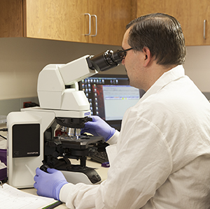
Lab tests on biopsy tissue
Lab tests will be done on the sample of cells that was taken during your breast biopsy. From these tests, your healthcare provider will learn if the tumor needs certain hormones to grow. Tumors that need hormones to grow are called hormone-receptor positive (HR+). This is because they have receptors for either estrogen or progesterone.
Another test is done on the biopsy sample to see if the cancer cells make too much of the growth-promoting protein called the human epidermal growth factor receptor 2, or HER2. Tumors that make a lot of this protein are known as HER2-positive and tend to grow more quickly. Treatments that target HER2 can be used for these cancers.
There are other tests that can be done on the cancer cells to learn more about the cancer and the types of treatment that may be needed. These results are sent to your healthcare provider as your pathology report. Talk with your healthcare provider about your report and what the information on it means.
Imaging tests
-
Chest X-ray. A chest X-ray uses a small amount of radiation to create an image of tissues inside your body. This test can show if cancer has spread to the lungs. The test takes only a few minutes and won't cause any pain.
-
Breast MRI. This test uses large magnets and a computer to make detailed images of tissues in the breast. An MRI is sometimes used to help decide if just the tumor should be taken out (lumpectomy) or if the whole breast should be removed (mastectomy). During an MRI of your breast, you’ll lie on your stomach on a scanning table. The table has openings for each breast to keep them from being compressed during the test. The table slides into a tube-like machine that contains the magnet. After a series of images have been taken, you may be given a contrast through a needle in your arm. This helps your healthcare provider see the tumor. The contrast is not radioactive. More images are then taken. The entire session takes about 1 hour. MRIs are also used to see if cancer has spread outside of the breast to the spine and brain. For these images, you lie face up on a table inside a narrow cylinder while the machine creates images of your spine and brain.
-
CT scan. This test uses a series of X-rays and a computer to make detailed images of tissues inside the body. A CT scan can show things, such as enlarged lymph nodes, a swollen spleen, or pockets of infection in your organs. During the test, you lie still on a table as it slides through the center of the ring-shaped CT scanner. Then the scanner sends a beam of X-rays at your body. You may be asked to hold your breath once or more during the scan. You may be asked to drink a contrast after the first set of pictures is taken. This contrast can help better show abnormal areas in your body. The contrast will pass out of your body over the next day or so through your bowel movements. If you have contrast through an IV in your arm, this may cause a feeling of warmth in your body for a few minutes. In rare cases, it can also cause hives or other allergic reactions. Tell the test technician if you don’t feel well during the test.
-
Ultrasound. This test uses sound waves and a computer to create images of the breast and axilla (underarm).
-
Bone scan. This test shows whether cancer has spread to your bones. For this test, you’ll be injected with a small amount of radioactive substance. This substance will travel through your bloodstream and collect in areas of abnormal bone growth. Pictures are taken with a special camera and any spots that show up may be caused by cancer. Spots may also light up due to things like arthritis or infection and will need to be investigated further. The bone scan is the most sensitive method available for showing breast cancer that has spread to the bones.
-
PET scan. A PET scan is used to find cancer cells in the body. A needle is used to put a radioactive sugar into a vein. The sugar travels through the blood throughout the body and is taken up by the cancer cells. Cancer cells are more active and use more sugar than normal cells usually do. A scanner is then used to get pictures of the body that contain the radioactive sugar. A PET scan combined with a CT scan is called a PET/CT.
Getting your test results
Your healthcare provider will talk with you about which tests you'll have. Make sure to get ready for the tests as directed. Ask questions and talk about any concerns you have.
Online Medical Reviewer:
Donna Freeborn PhD CNM FNP
Online Medical Reviewer:
Jessica Gotwals RN BSN MPH
Online Medical Reviewer:
Todd Gersten MD
Date Last Reviewed:
1/1/2023
© 2024 The StayWell Company, LLC. All rights reserved. This information is not intended as a substitute for professional medical care. Always follow your healthcare provider's instructions.